|
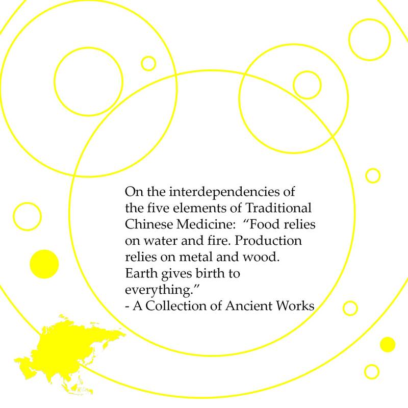
On the interdependencies of the five elements of Traditional Chinese Medicine: “Food relies on water and fire. Production relies on metal and wood. Earth gives birth to everything.”.- A Collection of Ancient Works
In Traditional Chinese Medicine connective tissue (including ligaments, tendons, spinal and brain's dura and meningis, carpal tendons, bones, pericardium (connective tissue sac around the heart, the protective layers around muscles and nerves, and more) is associated with Earth elements (stomach and spleen) as well as the mouth, the taste of sweetness and a sense of anxiety. One way to use this information is to do visualization on the color yellow which is also associated with the Earth elements. While resting quietly place one hand over the stomach (lower ribcage slightly to the left of center) and the other hand on the spleen (left side of the body, deep to the lower rib cage). Focus on your breathing and the color yellow. Imagine all the shades of yellow you can and all the things, clothes, birds, books, etc around you that are yellow. Another exercise is while you are walking or a passenger in a car, look around and notice everything you see that is yellow.
0 Comments
Connective Tissue Disorders and Manual Therapy
Manual therapy and Massage therapy practitioners, Acupuncturist, Integrative Manual Therapists, Osteopaths and Chiropractors use a variety of techniques to increase circulation and fluid flow, decrease scar tissue and pain, improve joint mobility and strength, reduce depression and eliminate the signs and symptoms of connective tissue and extracellular matrix disorders in the field of fasciology. They treat clients with a variety of connective tissue dysfunctions including: Benign Joint Hypermobility, Marfan’s syndrome, Ehlers-Danlos syndrome / Cutis Hyperelastica, osteogenesis imperfecta, Scleroderma, Rheumatoid arthritis, Raynaud's phenomenon, mixed connective tissue disease (MCTD), Cardiovascular disorders, Down’s syndrome, metabolic disorders (homocystinuria and hyperlysinemia), Fibromyalgia, Recurrent joint dislocations, Chronic low back pain, Carpal tunnel syndrome and Motor vehicle accident (MVA) injuries. This issue of The Burnham Review also focuses on the work on Helene Langevin, MD at the University of Vermont Medical School. She describes both the cellular properties of connective tissue and the effects of mechanical stretching and acupuncture in relationship to connective tissue. Many of the articles published by Dr. Langevin are available in full text. In a recent article, Langevin notes, “a patient presenting with a flareup of ulcerative colitis preceded by a two week exacerbation of knee osteoarthritis would probably be thought to have two distinct problems, one in the gut and one in the knee. Establishing the presence of a connective tissue ‘‘bridge’’ between these two medical problems would potentially have important repercussions on both diagnosis and treatment of these conditions.” 1. Langevin, H. M. (2006). "Connective tissue: a body-wide signaling network?" Med Hypotheses 66(6): 1074-7 [Full Text] www.anatomytrains.com/uploads/rich_media/c04c7252238ff6f72d81f52c89a20f85.pdf and Anatomy Trains http://www.anatomytrains.com/at/library/articles and Fascia Research Congress http://www.fascia2007.com/presenters.php. [Other Material on Connective Tissue Healing] https://www.nervewhisperer.solutions/peace/category/connective-tissue-healing Self-Supervision System of Fasciology The fascial network constituted by the connective tissues may be the anatomical basis for acupuncture therapy. Researchers found “acupoints were mainly located where thick connective tissues were present. In this fascial network, sensitive nerve endings, active cells and lymphatic vessels abounded in the sites with thick connective tissue, and needling at these sites induced definite biological effects. In light of biological phylogeny and embryo development, we believe that the connective tissue network may constitute a new functional system in the human body, the Self-supervision and control system.” 2. Wang, J., W. R. Dong, et al. (2007). "[From meridians and acupoints to self-supervision and control system: a hypothesis of the 10th functional system based on anatomical studies of digitized virtual human]." Nan Fang Yi Ke Da Xue Xue Bao 27(5): 573-9. http://www.camresearch.net/showabstract.php?pmid=17545059 and Wang, J., C. L. Wang, et al. (2007). "[Explanation of essence and substance basis of channels and collaterals with fasciology]." Zhongguo Zhen Jiu 27(8): 583-5. [PubMed Abstract] and [Abstract] http://www.camresearch.net/showabstract.php?pmid=17853756 Benign Joint Hypermobility "Benign joint hypermobility syndrome (BJHS) is a connective tissue disorder with hypermobility in which musculoskeletal symptoms occur in the absence of systemic rheumatologic disease. When patients with this syndrome are first seen by a physician, their chief complaint is joint pain, so BJHS can be easily overlooked. Treatment modalities include patient education, activity modification, stretching and strengthening exercises for the affected joint, and osteopathic manipulative treatment. Recurrent dislocation of the shoulder and patella as well as other orthopedic abnormalities are associated with joint laxity. Strength training should consist of a combination of both open kinetic (distal extremity moves freely) and closed kinetic chain (distal extremity meets resistance) exercises. Closed kinetic chain exercises often simulate functional demands, while open kinetic chain activities are better for more targeted strength training. Focused exercises to improve muscle strength, balance, and coordination may help improve joint stability and proprioception. Improvement of proprioception may reduce strain to the ligaments surrounding the joint and avoid further injury. Thrust treatment techniques [Osteopathic Manipulative Treatment] applying high velocity/low amplitude forces are the most widely used, but because of the increased tissue fragility seen in BJHS and weak supporting structures of the joint, gentler techniques like facilitated positional release and Counterstrain are good alternatives. OMT helps induce articular release resulting in increased joint motion, and reduced pain as well as improved blood flow, lymphatic drainage, and proprioception.” 3. Simpson, M. R. (2006). "Benign joint hypermobility syndrome: evaluation, diagnosis, and management." J Am Osteopath Assoc 106(9): 531-6. [Full Text] http://www.jaoa.org/cgi/content/full/106/9/531 Neural Effect of Connective Tissue Massage on Fibromyalgia “Connective tissue massage (CTM) is a manipulative technique that facilitates the diagnosis and treatment of a wide range of pathologies. Observation and subsequent manipulation of the skin and subcutaneous tissues can have a beneficial effect upon tissues remote from the area of treatment. These effects appear to be mediated by neural reflexes that cause an increase in blood flow to the affected region together with suppression of pain.” 4. Goats, G. C. and K. A. Keir (1991). "Connective tissue massage." Br J Sports Med 25(3): 131-3. [PubMed Abstract] and [Abstract] http://www.camresearch.net/showabstract.php?pmid=1777777 5. Michalsen, A. and M. Buhring (1993). "[Connective tissue massage]." Wien Klin Wochenschr 105(8): 220-7 [PubMed Abstract] [Abstract] http://www.camresearch.net/showabstract.php?pmid=8506683. Another randomized study of 48 individuals diagnosed with fibromyalgia showed “a series of 15 treatments with connective tissue massage conveys a pain relieving effect of 37%, reduces depression and the use of analgesics, and positively effects quality of life. The treatment effects appeared gradually during the 10-week treatment period.“ 6. Brattberg, G. and European Federation of Chapters of the International Association for the Study of Pain (1999). "Connective tissue massage in the treatment of fibromyalgia." Eur J Pain 3(3): 235-244 [PubMed Abstract] [Abstract] http://www.camresearch.net/showcitationlist.php?keyword=connective%20tissue%20fibroblasts. Osteopath, Andrew Taylor Still's View of Connective Tissue Dr Still “not only reset his patient’s hips, he also successfully treated goiters and acute appendicitis using manual techniques. He worked with muscles, bones, and joints, but, beyond that, “he worked with the fascia, which he described as the dwelling place of the soul.”7 (Still,1902). From a fascial perspective, muscles, bones, and joints are all included within the connective tissue. 7. Still, A. T. (1902). The Philosophy and Mechanical Principles of Osteopathy. Kansas City, Mo, Hudson-Kimberly Publication Co. [Full Text] 322 page e-book http://www.interlinea.org/atstill/eBookPMPO_V2.0.pdf. The extracellular matrix, the microscopic aspect of connective tissue, is inherently a gel. It is only because of the movement of calcium ions (ie, calcium waves) that nutrients and waste products flow to and from the cells. Calcium ions depolymerize and make watery this otherwise unyielding gel. Similarly, when osteopathic manipulative treatment is used to treat patients with somatic dysfunction, it decongests the connective tissues to restore their acidic, gel-like character to a healthy, fluid quality. Then blood, lymph, and nerve function can operate efficiently. Dr Still knew that congestion is antithetical to life when he said, fascia, “the framework of life,” is where we live and die. (Still,1902). According to Still because “anyone can find disease,” the goal of osteopathic medicine is to “find health” in the tissues. This goal means practitioners must look for ways to support healthy metabolic activity of the connective tissues in which cells reside.8 (Lee,2006). 8. Lee, R. P. (2006). "Still's concept of connective tissue: lost in "translation"?" J Am Osteopath Assoc 106(4): 176-7; author reply 213-4. [Full Text] http://www.jaoa.org/cgi/content/full/106/4/176 and Sutherland, W. G., DO and D. Ada Strand and Anne Wales, editors, (1998). Contributions of Thought: the Collected Writings of W.G. Sutherland. Fort Worth, Tex, Sutherland Cranial Teaching Foundation; 1998 [Reference] http://www.osteopathic.org/pdf/pub_osteolit.pdf and Rogers, F. J., G. E. D'Alonzo, Jr., et al. (2002). "Proposed tenets of osteopathic medicine and principles for patient care." J Am Osteopath Assoc 102(2): 63-5 [Full Text] http://www.jaoa.org/cgi/reprint/102/2/63. Extracellular Matrix Unifying Structure and Function Describing connective tissue as “Osteopathic Tissue” or the place where manual therapy techniques take place, Lee notes, “the extracellular matrix and the cytoskeleton operate as an unified electromechanical and chemomechanical system to turn cell functions on and off. All the necessary elements for the health and maintenance of the organism exist in, and pass through, the extracellular matrix. Thus, we can say that the connective tissues are holistic, demonstrating a structure function interrelationship and containing all the necessary resources for self-healing." 9. Lee, R. P. (2006). "Still's concept of connective tissue: lost in "translation"?" J Am Osteopath Assoc 106(4): 176-7; author reply 213-4. [Full Text] http://www.jaoa.org/cgi/content/full/106/4/176 Ehlers-Danlos, Osteogenesis Imperfecta and Manual Therapy Ehlers-Danlos Syndrome is a genetically based disorder, where the connective tissue is looser or more lax than it should due to problems with collagen synthesis. EDS is part of a group of connective tissue disorders characterized by abnormalities of the skin, ligaments and internal organs. The skin and blood vessels are extremely fragile and elastic. The skin is soft with rubber consistency and easily bruising. There are hypermobile joints with increased extensibility. There are also sometimes respiratory related problems. 10. Lopes, C., A. Manique, et al. (2006). "[Ehlers-Danlos syndrome - a rare cause of spontaneous pneumothorax]." Rev Port Pneumol 12(4): 471-80. [PubMed Abstract] http://www.ncbi.nlm.nih.gov/entrez/query.fcgi?cmd=Retrieve&db=PubMed&dopt=Citation&list_uids=16969576 “Orthopaedic complications such joint pain, joint swelling, joint dislocation, back pain, with walking and hand function disability are the main problems in Ehlers-Danos syndrome. Physical therapy has an important place in management." 11. Le Tallec, H., A. Lassalle, et al. (2006). "[Two cases of rehabilitation in Ehler-Danlos syndrome]." Ann Readapt Med Phys 49(2): 81-4. [PubMed Abstract] http://www.ncbi.nlm.nih.gov/entrez/query.fcgi?cmd=Retrieve&db=PubMed&dopt=Citation&list_uids=16430988 “Collagen mutations are directly responsible for the bone fragility of Osteogenesis Imperfecta and indirectly responsible for Ehlers-Danlos syndrome symptoms, by interference with N-propeptide removal.” 12. Cabral, W. A., E. Makareeva, et al. (2005). "Mutations near amino end of alpha1(I) collagen cause combined osteogenesis imperfecta/Ehlers-Danlos syndrome by interference with N-propeptide processing." J Biol Chem 280(19): 19259-69. [Full Text] http://www.jbc.org/cgi/content/full/280/19/19259 Integrative Manual Therapy and other hands-on techniques are used to help restore function and quality of life for people with Ehlers-Danlos syndrome, Osteogenesis Imperfecta and other connective tissue disorders which can lead to joint problems especially in the weight bearing joints of the legs. CAM Treatment for Scleroderma “Scleroderma is an autoimmune disease of the connective tissue characterized by fibrosis and thickening of various tissues. It can be limited to the skin or affect multiple organs, and its course ranges from slowly to rapidly progressive. There are “several promising natural treatments for scleroderma, including para-aminobenzoic acid, vitamin E, vitamin D, evening primrose oil, estriol, N-acetylcysteine, bromelain, and an avocado/soybean extract.” 13. Gaby, A. R. (2006). "Natural remedies for scleroderma." Altern Med Rev 11(3): 188-195. [Full Text] http://www.ncbi.nlm.nih.gov/entrez/query.fcgi?cmd=Retrieve&db=PubMed&dopt=Citation&list_uids=17217320 Acupuncture and Tissue Planes Helene Langevin, MD notes, “Acupuncture meridians traditionally are believed to constitute channels connecting the surface of the body to internal organs.” University of Vermont medical researchers, “found an 80% correspondence between the sites of acupuncture points and the location of intermuscular or intramuscular connective tissue planes in postmortem tissue sections.” Dr. Langevin continues, “because the structure and composition of interstitial connective tissue is responsive to mechanical stimuli, we propose that it plays a key role in the integration of several physiological functions with ambient levels of mechanical stress.” 14. Langevin, H. M. and J. A. Yandow (2002). "Relationship of acupuncture points and meridians to connective tissue planes." Anat Rec 269(6): 257-65. [Full Text] http://www.ncbi.nlm.nih.gov/entrez/query.fcgi?cmd=Retrieve&db=PubMed&dopt=Citation&list_uids=12467083 and [Full Text] http://www.med.uvm.edu/neurology/downloads/Relationshipofacupuncturepointsandmeridianstoconnectivetissueplanes.pdf IMT and Soft Tissue Injuries Integrative Manual Therapy (IMT) is being used to recovery function and decrease pain. A 2005 article describes some of the benefits. A car accident left Dina Drits with soft-tissue damage in her lower back and her pelvis out of alignment. “It hurt to brush my teeth and I couldn't walk for more than 15 minutes at a time. I couldn't do any of my regular sports and I was a ballroom dancer." After IMT treatment Drits reported, “I absolutely do things I couldn't do. Yoga and tai chi are both therapeutic. I can go on long hikes now." 15. Collins, L. (2005). "Healing hands Integrated manual therapy is alternative method of relieving pain." 2005(Sept 17): [Full Text] http://deseretnews.com/dn/view/0,1249,600108104,00.html. In an article aimed at helping athletes improve function, Holt writes, “manual therapies are time-consuming in the light of the simple task of hooking an athlete to a therapeutic machine. However, they are recognized as important techniques in controlling pain, restoring normal range of motion, and treating specialized conditions such as myofascial pain syndrome.” 16. Holt, J. (2004). "Manual Therapy and Athletic Injury Rehabilitation: Benefits of a Class of Therapy." The Sport Supplement, A Supplement of the Sports Journal Volume 12, Number 3: Summer(from www.thesportjournal.org/sport-supplement/vol12no3/03manual_therapy.asp and /www.friidrott.se/veteran/dokument/dunton/2004/trainin32.html). Holt lists the benefits of manual therapies in connective tissue, muscle and joint related problems: Massage Therapy for –> relief of spasm, increased lymphatic drainage, increased cutaneous circulation, increased cell metabolism, increased venous flow, increased extensibility of connective tissues, increased pliability of scar tissue, decreased neuromuscular excitability. Myofascial Release for –> relief of spasm, decrease in gamma gain/relaxation of hypersensitivity to stretch, relaxation of tight fascia, joint mobilization, restoration of correct joint function, stimulation of joint receptors, increased large-diameter afferent fiber input. Muscle Energy Technique for –> increased stretching of tight muscles/fascia, strengthening of weakened muscles, mobilization of restricted joints Strain-Counterstrain for –> reducing/arresting inappropriate proprioceptive activity 17. Holt, J. (2004). "Manual Therapy and Athletic Injury Rehabilitation: Benefits of a Class of Therapy." The Sport Supplement, A Supplement of the Sports Journal Volume 12, Number 3: Summer(from www.thesportjournal.org/sport-supplement/vol12no3/03manual_therapy.asp and /www.friidrott.se/veteran/dokument/dunton/2004/trainin32.html). The article also looks at the effects of Integrative Manual Therapy on knee pain. Lunn (2001) reported the case of an 18-year old male who had reconstructive surgery of the right anterior cruciate ligament following a skiing accident and subsequent re-injury. The patient used crutches, toe touch weight bearing only, no brace and medication for pain. The specific techniques used were Jones’ Strain-Counterstrain, lymph node and blood vessel Advanced Strain-counterstrain, Myofascial Release, Bone Bruise Therapy, Disruption of Membrane, and Neural Tissue Tension. Home exercises were also performed as directed by the physical therapist. After two days, there was significant improvement in quadriceps strength, ambulation, and range of motion in hip, knee, and ankle joints, as well as sensory improvement in the affected thigh.” 18. Holt, J. (2004). "Manual Therapy and Athletic Injury Rehabilitation: Benefits of a Class of Therapy." The Sport Supplement, A Supplement of the Sports Journal Volume 12, Number 3: Summer(from www.thesportjournal.org/sport-supplement/vol12no3/03manual_therapy.asp and /www.friidrott.se/veteran/dokument/dunton/2004/trainin32.html). Chiropractic Care Another article notes “two disabled patients diagnosed with Ehlers-Danlos syndrome had spinal pain, including neck and back pain, headache, and extremity pain. Commonalities among these 2 cases included abnormal spinal curvatures (kyphosis and scoliosis), joint hypermobility, and tissue fragility. One patient had postsurgical thoracolumbar spinal fusion (T11-sacrum) for scoliosis and osteoporosis. The other patient had moderate anterior head translation.” The clients both benefitted from joint mobilization techniques. Researchers noted, “both patients were able to reduce pain and anti-inflammatory medication usage in association with chiropractic care. Significant improvement in self-reported pain and disability as measured by visual analog score, Oswestry Low-Back Disability Index, and Neck Pain Disability Index were reported, and objective improvements in physical examination and spinal alignment were also observed following chiropractic care. Low-force chiropractic adjusting techniques may be a preferred technique of choice in patients with tissue fragility.” 19. Colloca, C. J. and B. S. Polkinghorn (2003). "Chiropractic management of Ehlers-Danlos syndrome: a report of two cases." J Manipulative Physiol Ther 26(7): 448-59.[PubMed Abstract] http://www.ncbi.nlm.nih.gov/entrez/query.fcgi?cmd=Retrieve&db=PubMed&dopt=Citation&list_uids=12975632 Manipulation and Sleep "The aim of the study was to evaluate the short-term and 1-year follow-up results of connective tissue manipulation of the back and combined ultrasound therapy (US and high-voltage pulsed galvanic stimulation) in terms of pain, complaint of nonrestorative sleep, and impact on the functional activities in patients with fibromyalgia (FM). Statistical analyses revealed that pain intensity, impact of FM on functional activities, and complaints of nonrestorative sleep improved after the 20 treatment program." 20. Citak-Karakaya, I., T. Akbayrak, et al. (2006). "Short and long-term results of connective tissue manipulation and combined ultrasound therapy in patients with fibromyalgia." J Manipulative Physiol Ther 29(7): 524-8. [PubMed Abstract] and [Abstract] http://www.camresearch.net/showabstract.php?pmid=16949941 Knee Problems Two books explain in detail ways to use Integrative Manual Therapy to work with the connective tissue and extremity joints Dr. Sharon W. Giammatteo’s most recent book focuses exclusively on connective tissue and the use of Integrative Manual Therapy for connective tissue disorders. 21. Weiselfish-Giammatteo, S., J. B. Kain, et al. (2005). Integrative manual therapy for the connective tissue system : myofascial release. Berkeley, Calif., North Atlantic Books. In a 2000 article W. Giammatteo describes some of the things a therapist working with connective tissue and structural disorders affecting the knees should consider, “muscle spasm of muscles which move the pelvis, hip, knee and ankle joints can contribute to decreased joint mobility and ranges of motion. The iliacus, piriformis, adductors, hamstrings, quadriceps, and gastrocnemius muscles typically compromise knee function when they are in spasm. Often there is scarring and hypertrophy of the connective tissue in the region of the knee, especially the joint capsule, the medial and lateral collateral ligaments, the tendons of medial hamstrings and gastrocnemius, and the cartilage of the meniscus. Compromised knee joint space causes friction which results in breakdown of the hyaline cartilage. Osteoarthritis will possibly occur. Occasionally circulation is affected which may cause a deep, burning discomfort in and around the knee joint. The common vascular problems which affect knee circulation are impingements and stenosis of the femoral artery and femoral vein at the hip joint region. The tibial lymph node which is inferior to the knee joint may be swollen secondary to infection, which causes pain, swelling and congestion of the knee and distal leg. Often knee joint problems may be secondary to limited dorsiflexion of the ankle joints: the tibiotalar and subtalar joints. When dorsiflexion is less than 10 degrees, the tibia cannot glide forward on talus during walking and running. This limitation of motion results in excessive extensor forces transcribed up the leg. In this scenario, the quadriceps goes into spasm, compresses the patella against the femur, causing a breakdown of hyaline cartilage behind the patella, causing chondromalasia.” 22. Giammatteo, S. (2000). What is Integrative Manual Therapy and How Does it Relate to Knee Injuries: from http://www.centerimtboulder.com/sportsinjuries_article1.htm. She goes on to say, “many approaches are utilized to affect healing on a cellular and also on a systems level. Improved structural integrity will change the status quo for improved potential. Functional Rehabilitation is then more effective.” 23. Giammatteo, S. (2000). What is Integrative Manual Therapy and How Does it Relate to Knee Injuries: from http://www.centerimtboulder.com/sportsinjuries_article1.htm. Homotoxicology’s Matrix “According to homotoxicology illness is defined as an overload of the connective tissue matrix with toxic substances, the homotoxins. In order to support elimination of these homotoxins, complex homeopathic medicines were developed. Fibroblasts are the local cells of matrix and produce and modulate the composition of the extracellular matrix (ECM) in every organ. As the modulation of the ECM is dependent on the activity of the fibroblasts, plant extracts may modulate the composition of the ECM via the inhibitory effect on fibroblasts cell growth.” 24. Valentiner, U., M. Weiser, et al. (2003). "The effect of homeopathic plant extract solutions on the cell proliferation of human cutaneous fibroblasts in vitro." Forsch Komplementarmed Klass Naturheilkd 10(3): 122-7. [PubMed Abstract] http://www.ncbi.nlm.nih.gov/entrez/query.fcgi?cmd=Retrieve&db=PubMed&dopt=Citation&list_uids=12853718 Connective Tissue Dysplasia and Terri G Varicosities An analysis was made of two groups of patients presenting with varicosities. The first group comprised 82 patients aged 15 to 30 years without risk factors. The second group accrued 85 patients with traditional risk factors: pregnancy and birth, overweight, considerable dynamic and physical loading, age from 30 to 50 years, and intake of hormonal contraceptives. It has been established that the key role in the development of varicosis is played by connective tissue dysplasia (CTD), the intensity of which predetermines the origination of phlebopathy and varicosis as well as the rate of their progression. The most frequently occurring is the mechanism of the development of phlebopathy as structural and functional defectiveness of all venous vessels of the extremity, leading to the rise of the deposited blood volume in the leg because of the high elasticity of venous walls. Secondly, CTD that initially impairs valve morphology, results in local varicosis under the effect of reflux hydrodynamic strokes at the weakened venous wall.” 25. Tsukanov Iu, T. and A. Tsukanov (2004). "[Varicosis of the lower extremities as a consequence of connective tissue dysplasia]." Angiol Sosud Khir 10(2): 84-9. http://www.ncbi.nlm.nih.gov/entrez/query.fcgi?cmd=Retrieve&db=PubMed&dopt=Citation&list_uids=15163975 Systemic Atherosclerosis "A 47-year-old woman presented with facial spasm, swollen fingers and Raynaud's phenomenon due to cerebrovascular disorder and mixed connective tissue disease (MCTD). In this case, systemic atherosclerosis might have been linked to these autoimmune reactions." 26. Kanazawa, M., Y. Wada, et al. (2004). "Mixed connective tissue disease associated with antineutrophil cytoplasmic antibodies against proteinase-3 and systemic atherosclerosis: a case report." Clin Rheumatol 23(5): 456-9. [PubMed Abstract] http://www.ncbi.nlm.nih.gov/entrez/query.fcgi?cmd=Retrieve&db=PubMed&dopt=Citation&list_uids=15459817 Blood Vessel Walls in Ehlers-Danlos and Nutrition Problems of the blood vessel wall integrity and easy bruising is a common feature in Ehlers-Danlos syndrome. 27. Yen, J. L., S. P. Lin, et al. (2006). "Clinical features of Ehlers-Danlos syndrome." J Formos Med Assoc 105(6): 475-80. There are manual therapy techniques to address membrane wall fragility (Disruption of Membrane technique). 28. Weiselfish-Giammatteo, S. (2000) Disruptions of Membrane. from www.centerimt.com/Products/videos.asp. Bloomfield, CT, Dialogues in Contemporary Rehabilitation. People should also consider the benefits of essential fatty acids (fish oils) and antioxidants in the healing of membrane wall weakness. Smooth Muscle in Fibroblasts “Alpha smooth muscle actin was recently shown to be present in mouse subcutaneous tissue fibroblasts in the absence of tissue injury. 29. Storch, K. N., D. J. Taatjes, et al. (2007). "Alpha smooth muscle actin distribution in cytoplasm and nuclear invaginations of connective tissue fibroblasts." Histochem Cell Biol 127(5): 523-30. [PubMed Abstract] http://www.ncbi.nlm.nih.gov/entrez/query.fcgi?cmd=Retrieve&db=PubMed&dopt=Citation&list_uids=17310383 Green Tea, Essential Fatty Acids “Some green tea catechins are chondroprotective. Consumption of green tea may be prophylactic for arthritis and may benefit the arthritis patient by reducing inflammation.” 30. Adcocks, C., P. Collin, et al. (2002). "Catechins from green tea (Camellia sinensis) inhibit bovine and human cartilage proteoglycan and type II collagen degradation in vitro." J Nutr 132(3): 341-6. [Full Text] http://jn.nutrition.org/cgi/content/full/132/3/341 Essential fatty acids, especially polyunsaturated fatty acids (PUFA) can benefit cartilage and connective tissue disorders. One study found, “the pathologic indicators manifested in human OA cartilage can be significantly altered by exposure of the cartilage to n-3 PUFA, but not to other classes of fatty acids.” 31. Curtis, C. L., S. G. Rees, et al. (2002). "Pathologic indicators of degradation and inflammation in human osteoarthritic cartilage are abrogated by exposure to n-3 fatty acids." Arthritis Rheum 46(6): 1544-53. [Full Text] retracted http://www.ncbi.nlm.nih.gov/entrez/query.fcgi?cmd=Retrieve&db=PubMed&dopt=Citation&list_uids=12115185 Another study found, “n-3 PUFA (represented by EPA in this study) positively affect the healing characteristics of medial collateral ligament (MCL) cells and therefore may represent a possible noninvasive treatment to improve ligament healing.” 32. Hankenson, K. D., B. A. Watkins, et al. (2000). "Omega-3 fatty acids enhance ligament fibroblast collagen formation in association with changes in interleukin-6 production." Proc Soc Exp Biol Med 223(1): 88-95. [Full Text] http://www.ebmonline.org/cgi/content/full/223/1/88 Low Level Laser / Cold Laser Low level laser therapy (LLLT) is a therapeutic intervention used by manual therapist for back pain. “Low level laser therapy is a non-invasive light source treatment that generates a single wavelength of light. It emits no heat, sound, or vibration. It is also referred to as photobiology or biostimulation. LLLT is believed to affect the function of connective tissue cells (fibroblasts), accelerate connective tissue repair and act as an anti-inflammatory agent. Lasers with different wavelengths, varying from 632 to 904 nm, are used in the treatment of musculoskeletal disorders. 33. Yousefi-Nooraie, R., E. Schonstein, et al. (2007). "Low level laser therapy for nonspecific low-back pain." Cochrane Database Syst Rev(2): CD005107. [Abstract] http://mrw.interscience.wiley.com/cochrane/clsysrev/articles/CD005107/frame.html 34. Luminex Medical Laser System 4 different heads with laser diodes of an average power of 500mW at a wavelength of 867nm[Full Text] www.medicallasersystems.com Infertility and Manual Therapy “In a study to assess the effectiveness of site-specific manual soft tissue therapy in facilitating natural fertility and improving in vitro fertilization (IVF) pregnancy rates in women with histories indicating abdominopelvic adhesion formation, researchers found, "this innovative site-specific protocol of manual soft-tissue therapy facilitates fertility in women with a wide array of adhesion-related infertility and biomechanical reproductive organ dysfunction. The therapy, designed to improve function by restoring visceral, osseous, and soft-tissue mobility, is a nonsurgical, noninvasive manual technique with no risks and few, if any, adverse side effects or complications. Mobilization of the soft tissues using site-specific manual therapy appears to break the attachments of the collagenous cross-links within the adhesions, thus restoring normal mobility and function to the previously adhered organs. According to Mojzisovà, "there is a direct relationship between the condition of the lower back muscles and the way the reproductive organs function." 35. Mojzis L, Nemec R, Hlavaty V. Children of Your Own: the Mojzis Method. Boulder, Colo: Richmond Bay; 1990. and (Volejnikova,2001). Volejnikova H. Female infertility: a study of physical treatment by the method of L. Mojzisovà for functional disturbances of the pelvic region. J Orthopaedic Med. 2001;23:47-49. The purpose of the second Prague study, based on 2006 randomly selected infertile women, was to determine which types of infertility were best suited for treatment by the Mojzisovà method. Results showed that conception rates ranged from a low of 11% for women aged 40 to 44 to a high of 46% for the age group 20 to 24. 36. Wurn, B. F., L. J. Wurn, et al. (2004). "Treating Female Infertility and Improving IVF Pregnancy Rates With a Manual Physical Therapy Technique." Medscape General Medicine 6(2):51: [Full Text] http://www.medscape.com/viewarticle/480429_print and www.clearpassage.com. Back Pain Mechanism and Fear "We hypothesize that pain-related fear leads to a cycle of decreased movement, connective tissue remodeling, inflammation, nervous system sensitization and further decreased mobility. The integration of connective tissue and nervous system plasticity into the model will potentially illuminate the mechanisms of a variety of treatments that may reverse these abnormalities by applying mechanical forces to soft tissues (e.g. physical therapy, massage, chiropractic manipulation, acupuncture), by changing specific movement patterns (e.g. movement therapies, yoga) or more generally by increasing activity levels (e.g. recreational exercise). An integrative mechanistic model incorporating behavioral and structural aspects of cLBP will strengthen the rationale for a multidisciplinary treatment approach including direct mechanical tissue stimulation, movement reeducation, psychosocial intervention and pharmacological treatment to address this common and debilitating condition." 37. Langevin, H. M. and K. J. Sherman (2007). "Pathophysiological model for chronic low back pain integrating connective tissue and nervous system mechanisms." Med Hypotheses 68(1): 74-80. [Full Text] www.chiro.org/LINKS/ABSTRACTS/Pathophysiological_Model_for_Chronic_Low_Back_Pain.shtml[PubMed Abstract] www.ncbi.nlm.nih.gov/entrez/query.fcgi?cmd=Retrieve&db=PubMed&dopt=Citation&list_uids=16919887 Chiropractor, Dan Murphy, highlighted a number of key points: 1) In chronic low back pain, there is an integration between connective tissue fibrosis and the nervous system perception of pain. 2) Adverse connective tissue fibrosis can be remodeled by applying mechanical forces to soft tissues, (chiropractic spinal adjusting). 3) The association between symptoms and imaging results (X-ray, CT, MRI) has been consistently weak, and up to 85 percent of patients with low back pain cannot be given a precise pathoanatomical diagnosis. 4) Ongoing pain is associated with widespread neuroplastic changes at multiple levels within the nervous system, including primary afferent neurons, spinal cord, brainstem, thalamus, limbic system and cortex." 5) There are distinct "brain networks" involved in acute vs. chronic pain. Chronic pain is specifically related to regions for cognition and emotions. 6) Chronic back pain results in neuronal or glial loss in the pre-frontal and thalamic gray matter. 38. Dr. Dan Murphy,DC from [Full Text]www.chiro.org/LINKS/ABSTRACTS/Pathophysiological_Model_for_Chronic_Low_Back_Pain.shtml According to Langevin & Sherman, evidence supports the fact that among chronic low back patients, pain affects how they move, resulting in abnormal trunk muscle activity during postural perturbation, impaired control of trunk and hip during arm movements and abnormal postural compensation for respiration. As a result of emotional, behavioral and motor dysfunction, abnormal connective tissue remodeling, inflammation, nervous system sensitization and further decreased mobility occurs, creating a vicious cycle." 39. Warren, H (2006) Fibrosis May Be Related to Chronic Pain www.warrenhammer.comand www.chiroweb.com/columnist/hammer Fibrosis May Be Related to Chronic Pain Langevin and Sherman stressed the association of abnormal connective tissue with the nervous system: "Both increased stress due to overuse, repetitive movement and / or hypermobility, and decreased stress due to immobilization or hypomobility can cause changes in connective tissue." Both hyper- and hypomobility can result in either atrophy or fibrosis. Inflammation, tissue hypo-oxygenation and cytokines such as TGF-1 will promote fibrosis. Whether it is the presence of trigger points within the fascia causing painful muscle contraction, microinjury, inflammation, growth factors or abnormal biomechanics, there will be an increase in fibrosis, leading to increased tissue stiffness and further loss of motion. “They also state that research into this area will help explain why treatments such physical therapy, massage, chiropractic manipulation, acupuncture, movement therapies and yoga may be valuable.” 40. Warren, H (2006) Fibrosis May Be Related to Chronic Pain www.warrenhammer.comand www.chiroweb.com/columnist/hammer Neck and Dura Connection "The connective tissue attachments to the cervical spinal dura mater originate from the ligamentum nuchae (LN) and rectus capitis posterior minor (RCPM) muscle Our results indicate that: 1) the attachments between the LN and RCPM and the dura occur between vertebrae C1-C2 and the occipital bone and C1, respectively, and that they are substantial normal anatomic attachments, 2) attachments between the LN and RCPM are usually present, and 3) the attachments between the LN and dura mater can be identified on MRI. These latter attachments may play a role in neck pain, making their MRI appearance clinically important." 41. Humphreys, B. K., S. Kenin, et al. (2003). "Investigation of connective tissue attachments to the cervical spinal dura mater." Clin Anat 16(2): 152-9. [PubMed Abstract] and [Abstract] http://www.camresearch.net/showabstract.php?pmid=12589671 Electrical Properties “Acupuncture points and meridians are commonly believed to possess unique electrical properties. Recent studies indicate a correspondence between acupuncture meridians and connective tissue planes. We hypothesized that segments of acupuncture meridians that are associated with loose connective tissue planes (between muscles or between muscle and bone) visible by ultrasound have greater electrical conductance (less electrical impedance) than non-meridian, parallel control segments. Meridian segments were determined by palpation and proportional measurements. Connective tissue planes underlying those segments were imaged with an ultrasound scanner. Along each meridian segment, four gold-plated needles were inserted along a straight line and used as electrodes. A parallel series of four control needles were placed 0.8 cm medial to the meridian needles. Tissue impedance was on average lower along the Pericardium meridian, but not along the Spleen meridian, compared with their respective controls. Ultrasound imaging of meridian and control segments suggested that contact of the needle with connective tissue may explain the decrease in electrical impedance noted at the Pericardium meridian. 42. Ahn, A. C., J. Wu, et al. (2005). "Electrical impedance along connective tissue planes associated with acupuncture meridians." BMC Complement Altern Med 5: 10. [Full Text] http://www.pubmedcentral.nih.gov/articlerender.fcgi?tool=pubmed&pubmedid=15882468 Energy and TCM "There are three inter-related levels of a macromolecular energy-information relay system in the human body, each generated by a specific type of semi-conductant tissue and each with a specific function. The surface layer of the energy body, generated by fluid connective tissue and known as the ordinary channel system or meridian system in traditional Chinese medicine (TCM), functions in the service of immunosurveillance through detection of distress signals and transmitting energy-information regarding immunoresponse. The middle layer of the energy body, generated by semi-conductant hard and spongy bone tissue, known as the extraordinary channel system in TCM, functions in the service of longevity and regeneration, as described in Bodhidharma's classic, Bone Marrow Washing. The bone marrow energy-information system has direct relevance to modern stem cell research on the role of stem cells in regeneration of injured tissue. The deepest layer of the nery body, generated by semi-conductant nervous system tissue, notably the vagus nerve and spinal column, functions in the services of awakening consciousness and in immortality. This system is described in the Tibetan Inner Fire meditations as well as in the Taoist shen breathing practices. There is very little scientific understanding of the central channel system." 43. Brown, D. P. (2007). "The Energy Body and Its Functions: Immunosurveillance, Longevity, and Regeneration." Ann N Y Acad Sci. [Abstract] http://www.camresearch.net/showabstract.php?pmid=17905935 Acupucnture “Acupuncture needle rotation has been previously shown to cause specific mechanical stimulation of subcutaneous connective tissue. Needle rotation induced extensive fibroblast spreading and lamellipodia formation within 30 min, measurable as an increased in cell body cross sectional area. Significant effects of rotation were present throughout the tissue, indicating the presence of a response extending laterally over several centimeters.” 44. Langevin, H. M., N. A. Bouffard, et al. (2006). "Subcutaneous tissue fibroblast cytoskeletal remodeling induced by acupuncture: evidence for a mechanotransduction-based mechanism." J Cell Physiol 207(3): 767-74. [Full Text] http://drwdowin.com/Research.html In many cell types such as fibroblasts, endothelial cells, and sensory neurons, focal adhesions form a mechanical link between extracellular collagen matrix and intracellular cytoskeleton. 45. Chicurel, M. E., Chen, C. S., Ingber, D. E. (1998) Cellular control lies in the balance of forces. Curr. Opin. Cell Biol. 10,232-239 46. Giancotti, F. G., Ruoslahti, E. (1999) Integrin signaling. Science 285,1028-1032 [Full Text] http://www.sciencemag.org/cgi/content/abstract/285/5430/1028?ijkey=61ec39623ca419e89a29e55d4dce4dad1d93edb6&keytype2=tf_ipsecsha The mechanism of mechanical load detection is thought to be a mechanosensory complex composed of extracellular matrix-integrin-cytoskeletal components linked to a kinase cascade. 47. Burridge, K., Fath, K., Kelly, T., Nuckolls, G., Turner, C. (1988) Focal adhesions: transmembrane junctions between the extracellular matrix and the cytoskeleton. Annu. Rev. Cell Biol. 4,487-525 In this model, load deformation displaces matrix molecules tethered to clustered integrins at focal adhesions. 48. Muller, J. M., Chilian, W. M., Davis, M. J. (1997) Integrin signaling transduces shear stress-dependent vasodilatation of coronary arterioles. Circ. Res. 80,320-326 [Full Text]http://circres.ahajournals.org/cgi/content/full/80/3/320?ijkey=5b507f251758363034d127ceb355c7babf84809a The cell membrane displacement is transduced by an integrin to an integrin binding protein such as talin and then to associated proteins such as vinculin, tensin, paxillin, Src, and focal adhesion kinase 49. Clark, E. A., Brugge, J. S. (1995) Integrins and signal transduction pathways: the road taken. Science 268,233-239 [Full Text] http://www.sciencemag.org/cgi/content/abstract/268/5208/233?ijkey=3ac4cbb92d7d33ab01005d747add96aa7b2c9c5f&keytype2=tf_ipsecsha In addition, one or more of these proteins can undergo a conformation change in response to displacement and initiate a series of phosphorylation and binding reactions in the protein complex. Therefore, the result of mechanical load deformation of an integrin molecule via extracellular matrix attachment is activation of a signaling cascade leading to a wide range of cellular responses, including changes in the actin cytoskeleton with formation of stress fibers. 50. Banes, A. J., Tsuzaki, M., Yamamoto, J., Fischer, T., Brigman, B., Brown, T., Miller, M. (1995) Mechanoreception at the cellular level: the detection, interpretation and diversity of responses to mechanical signals 51. Banes, A. J., Tsuzaki, M., Yamamoto, J., Fischer, T., Brigman, B., Brown, T., Miller, M. (1995) Mechanoreception at the cellular level: the detection, interpretation and diversity of responses to mechanical signals. Biochem. Cell Biol. 73,349-365. 52. Dartsch, P. C., Hammerle, H. (1986) Orientation response of arterial smooth muscle cells to mechanical stimulation. Eur. J. Cell Biol. 41,339-346 53. Sumpio, B. E., Banes, A. J., Buckley, M., Johnson, G. (1988) Alterations in aortic endothelial cell morphology and cytoskeletal protein synthesis during cyclic tensional deformation. J. Vasc. Surg. 7,130-138. (Sumpio,1988). The pulling of collagen fibers induced by acupuncture needle manipulation appears to have a similar effect on connective tissue fibroblasts via their attachment to collagen fibers at focal adhesion complexes. These observations suggest that the mechanical signal created by acupuncture needle manipulation can induce intracellular cytoskeletal rearrangements in fibroblasts and possibly in other cells present within connective tissue, such as capillary endothelial cells. Cytoskeletal reorganization in response to mechanical load signals has been shown to induce cell contraction, migration, and protein synthesis. Potentially powerful effects may derive from this mechanical signal transduction, including autocrine and paracrine cellular effects, with modification of the surrounding extracellular matrix. 54. Sumpio, B. E., Banes, A. J., Buckley, M., Johnson, G. (1988) Alterations in aortic endothelial cell morphology and cytoskeletal protein synthesis during cyclic tensional deformation. J. Vasc. Surg. 7,130-138 55. Harris, A. K., Wild, P., Stopak, D. (1980) Silicone rubber substrata: a new wrinkle in the study of cell locomotion. Science 208,177-179. [Full Text] http://www.sciencemag.org/cgi/content/abstract/208/4440/177?ijkey=3fc4fd3eee0db9f991198c90f73f8755d3dc832d&keytype2=tf_ipsecsha In summary, the insertion and manipulation of acupuncture needles may have both local and remote therapeutic effects based on the same underlying mechanism: mechanical coupling of needle to connective tissue, winding of tissue around the needle, generation of a mechanical signal by pulling of collagen fibers during needle manipulation, and mechanotransduction of the signal into cells. Downstream effects of this mechanical signal may include cell secretion, modification of extracellular matrix, amplification and propagation of the signal along connective tissue planes, and modulation of afferent sensory input via changes in the connective tissue milieu. 56. Langevin, H. M., D. L. Churchill, et al. (2001). "Mechanical signaling through connective tissue: a mechanism for the therapeutic effect of acupuncture." Faseb J 15(12): 2275-82. [Full Text] http://www.fasebj.org/cgi/content/abstract/15/12/2275 Mechanical Stretch Researchers reported, brief (10 min) static tissue stretch attenuated the increase in both soluble Transforming growth factor beta 1 (TGF-beta1) (ex vivo) and Type-1 procollagen (in vivo) following tissue injury. These results have potential relevance to the mechanisms of treatments applying brief mechanical stretch to tissues (e.g., physical therapy, respiratory therapy, mechanical ventilation, massage, yoga, acupuncture).” 57. Bouffard, N. A., K. R. Cutroneo, et al. (2007). "Tissue stretch decreases soluble TGF-beta1 and type-1 procollagen in mouse subcutaneous connective tissue: Evidence from ex vivo and in vivo models." J Cell Physiol. [PubMed Abstract] http://www.ncbi.nlm.nih.gov/entrez/query.fcgi?cmd=Retrieve&db=PubMed&dopt=Citation&list_uids=17654495 Ultrasound Shows Tissue Changes “The goal of this study was to show that ultrasound can be used to quantify dynamic changes in local connective tissue structure in vivo. 3-D renditions of ultrasound images showed longitudinal echogenic sheets that matched with collagenous sheets seen in histological preparations. The combination of ultrasound and semi-variogram analyses allows quantitative assessment of dynamic changes in the structure of human connective tissue in vivo.” 58. Langevin, H. M., D. M. Rizzo, et al. (2007). "Dynamic morphometric characterization of local connective tissue network structure in humans using ultrasound." BMC Syst Biol 1: 25. [Full Text] http://www.pubmedcentral.nih.gov/articlerender.fcgi?tool=pubmed&pubmedid=17550618 Connecting and Communicating “Unspecialized "loose" connective tissue forms an anatomical network throughout the body.” Langevin “hypothesis that, in addition, connective tissue functions as a body-wide mechanosensitive signaling network. Three categories of signals are discussed: electrical, cellular and tissue remodeling, each potentially responsive to mechanical forces over different time scales. It is proposed that these types of signals generate dynamic, evolving patterns that interact with one another. Such connective tissue signaling would be affected by changes in movement and posture, and may be altered in pathological conditions (e.g. local decreased mobility due to injury or pain). Connective tissue thus may function as a previously unrecognized whole body communication system. Understanding the temporal and spatial dynamics of connective tissue bioelectrical, cellular and tissue plasticity responses, as well as their interactions with other tissues, may be key to understanding how pathological changes in one part of the body may cause a cascade of ‘‘remote’’ effects in seemingly unrelated areas and organ systems.59. Langevin, H. M. (2006). "Connective tissue: a body-wide signaling network?" Med Hypotheses 66(6): 1074-7. [Full Text] www.anatomytrains.com/uploads/rich_media/c04c7252238ff6f72d81f52c89a20f85.pdf and Anatomy Trains http://www.anatomytrains.com/at/library/articles and Fascia Research Congress http://www.fascia2007.com/presenters.php Mechanical Stretching Mechanical stretching of connective tissue occurs with normal movement and postural changes, as well as treatments including physical therapy, massage and acupuncture. Connective tissue fibroblasts were recently shown to respond actively to short-term mechanical stretch (minutes to hours) with reversible cytoskeletal remodeling, characterized by extensive cell spreading and lamellipodia formation. In unstretched tissue, the pattern of alpha-actin was diffuse and granular. With tissue stretch (30 min), alpha-actin formed a star-shaped pattern centered on the nucleus, while beta-actin extended throughout the cytoplasm including lamellipodia and cell cortex. This dual response pattern of alpha- and beta-actin may be an important component of cellular mechanotransduction mechanisms relevant to physiologic and therapeutic mechanical forces applied to connective tissue.” 60. Langevin, H. M., K. N. Storch, et al. (2006). "Fibroblast spreading induced by connective tissue stretch involves intracellular redistribution of alpha- and beta-actin." Histochem Cell Biol 125(5): 487-95. [PubMed Abstract] http://www.ncbi.nlm.nih.gov/entrez/query.fcgi?cmd=Retrieve&db=PubMed&dopt=Citation&list_uids=16416024
Cellular Stretching & Tissue Shape
“Cytoskeleton-dependent changes in cell shape are well-established factors regulating a wide range of cellular functions including signal transduction, gene expression, and matrix adhesion. Although the importance of mechanical forces on cell shape and function is well established in cultured cells, very little is known about these effects in whole tissues or in vivo. Tissue stretch ex vivo (average 25% tissue elongation from 10 min to 2 h) caused a significant time-dependent increase in fibroblast cell body perimeter and cross-sectional area. At 2 h, mean fibroblast cell body cross-sectional area was 201% greater in stretched than in unstretched tissue. Fibroblasts in stretched tissue had larger, "sheetlike" cell bodies with shorter processes. In contrast, fibroblasts in unstretched tissue had a "dendritic" morphology with smaller, more globular cell bodies and longer processes. The dynamic, cytoskeleton-dependent responses of fibroblasts to changes in tissue length demonstrated in this study have important implications for our understanding of normal movement and posture, as well as therapies using mechanical stimulation of connective tissue including physical therapy, massage, and acupuncture. 61. Langevin, H. M., N. A. Bouffard, et al. (2007). "Connective tissue fibroblast response to acupuncture: dose-dependent effect of bidirectional needle rotation." J Altern Complement Med 13(3): 355-60. [Abstract] http://www.acupuncture.com/newsletters/m_jun07/res.htm#1 Loose connective tissue forms a continuous network throughout the body, including subcutaneous and interstitial connective tissues surrounding and permeating muscles and organs. Therapeutic mechanical deformation of loose connective tissue is used routinely in physiotherapy (e.g., in stress-relaxation techniques), as well as in many “alternative” therapies such as massage, myofascial release, and osteopathic and chiropractic manipulations. In addition, acupuncture was recently shown to cause winding, pulling, and deformation of subcutaneous connective tissue. Mechanotransduction through connective tissue with resultant effects on fibroblast cell shape and function was recently proposed as a mechanism for the therapeutic effect of acupuncture. Understanding the downstream cellular and molecular effects of mechanotransduction in loose connective tissue may therefore give key insights into the therapeutic mechanisms of a variety of treatments for musculoskeletal pain.” 62. Langevin, H. M., N. A. Bouffard, et al. (2005). "Dynamic fibroblast cytoskeletal response to subcutaneous tissue stretch ex vivo and in vivo." Am J Physiol Cell Physiol 288(3): C747-56. [Full Text] http://ajpcell.physiology.org/cgi/content/full/288/3/C747 and Langevin HM, Churchill DL, and Cipolla MJ. Mechanical signaling through connective tissue: a mechanism for the therapeutic effect of acupuncture. FASEB J 15: 2275–2282, 2001. and Langevin HM, Churchill DL, Wu J, Badger GJ, Yandow J, Fox JR, and Krag MH. Evidence of connective tissue involvement in acupuncture.FASEB J 16: 872–874, 2002. 63. Langevin, H. M., N. A. Bouffard, et al. (2007). "Connective tissue fibroblast response to acupuncture: dose-dependent effect of bidirectional needle rotation." J Altern Complement Med 13(3): 355-60. [Abstract] http://www.acupuncture.com/newsletters/m_jun07/res.htm#1 Molecular Mimicry, Pathogens and Connective Tissue Aggregatibacter (formerly Actinobacillus) actinomycetemcomitans64 (Ready,2007) is just one of the thousands of different types of bacterium found in the mouth and is one of the causes of gum disease. Not only does gum disease put teeth at risk, but it has also been linked with certain kinds of heart disease. The star-shaped pattern within the colony is typical of these bacteria when grown under suitable conditions. 64. Ready, Darren Wellcome (2007) [Image] http://www.wellcome.ac.uk/en/bia/gallery.html?image=6) 65. Tabeta, K., H. Yoshie, et al. (2001). "Characterization of serum antibody to Actinobacillus actinomycetemcomitans GroEL-like protein in periodontitis patients and healthy subjects." Oral Microbiol Immunol 16(5): 290-5. [PubMed Abstract] http://www.ncbi.nlm.nih.gov/entrez/query.fcgi?cmd=Retrieve&db=PubMed&dopt=Citation&list_uids=11555306 If these bacteria escape their normal habitats, the surface components that mimic the sLe(x) oligosaccharide might bind to host antigens of the selectin family which could promote binding to endothelial cells and, consequently, initiation of the events leading to infective endocarditis. 66. Hirota, K., H. Kanitani, et al. (1995). "Cross-reactivity between human sialyl Lewis(x) oligosaccharide and common causative oral bacteria of infective endocarditis." FEMS Immunol Med Microbiol 12(2): 159-64. [PubMed Abstract] http://www.ncbi.nlm.nih.gov/entrez/query.fcgi?cmd=Retrieve&db=PubMed&dopt=Citation&list_uids=8589666 In another study researchers looked at, “the adhesion of A. actinomycetemcomitans to extracellular matrix components of the connective tissue. Mutants were identified which exhibited the following phenotypes: a decrease in collagen binding; a decrease in fibronectin binding; a decrease in binding to both proteins; and an increase in binding to both collagen and fibronectin. Collectively, the results support the hypothesis that A. actinomycetemcomitans host colonization involves afimbrial adhesins for extracellular matrix proteins, and that the expression of adhesion is modulated by global regulatory mechanisms. 67. Mintz, K. P. (2004). "Identification of an extracellular matrix protein adhesin, EmaA, which mediates the adhesion of Actinobacillus actinomycetemcomitans to collagen." Microbiology 150(Pt 8): 2677-88. [Full Text] http://mic.sgmjournals.org/cgi/content/full/150/8/2677?view=long&pmid=15289564 68. Ruiz, T., C. Lenox, et al. (2006). "Novel surface structures are associated with the adhesion of Actinobacillus actinomycetemcomitans to collagen." Infect Immun 74(11): 6163-70. [Full Text] http://www.pubmedcentral.nih.gov/articlerender.fcgi?tool=pubmed&pubmedid=17057091 Extracellular Matrix The ECM is a biologically active tissue composed of a complex mixture of macromolecules, including multiple collagen types, fibronectin, laminin and glycosaminoglycans. The ECM not only serves a structural function but also affects a number of cellular activities, including migration, proliferation and differentiation. ECM proteins that have been described to act as a substrate for bacterial adhesion include collagens, laminin, fibronectin, fibrinogen, vitronectin and heparan sulfate. 69. Patti, J. M., Allen, B. L., McGavin, M. J. & Hook, M. (1994). MSCRAMM-mediated adherence of microorganisms to host tissues. Annu Rev Microbiol 48, 585–617. [PubMed Abstract] In addition, ubiquitous signaling molecules have also been identified that regulate the adhesion of this pathogen to these substrates.” 70. Mintz, K. P. (2004). "Identification of an extracellular matrix protein adhesin, EmaA, which mediates the adhesion of Actinobacillus actinomycetemcomitans to collagen." Microbiology 150(Pt 8): 2677-88. [Full Text] http://mic.sgmjournals.org/cgi/content/full/150/8/2677?view=long&pmid=15289564 Selected Work Helene M Langevin, MD et al. 1. Langevin, H. M., D. M. Rizzo, et al. (2007). "Dynamic morphometric characterization of local connective tissue network structure in humans using ultrasound." BMC Syst Biol 1: 25 [Full Text] http://www.pubmedcentral.nih.gov/articlerender.fcgi?tool=pubmed&pubmedid=17550618. 2. Langevin, H. M., N. A. Bouffard, et al. (2006). "Subcutaneous tissue fibroblast cytoskeletal remodeling induced by acupuncture: evidence for a mechanotransduction-based mechanism." J Cell Physiol 207(3): 767-74 [Full Text] http://drwdowin.com/Research.html. 3. Langevin, H. M. (2006). "Connective tissue: a body-wide signaling network?" Med Hypotheses 66(6): 1074-7 [Full Text] www.anatomytrains.com/uploads/rich_media/c04c7252238ff6f72d81f52c89a20f85.pdf and Anatomy Trains http://www.anatomytrains.com/at/library/articles and Fascia Research Congress http://www.fascia2007.com/presenters.php. 4. Langevin, H. M., N. A. Bouffard, et al. (2005). "Dynamic fibroblast cytoskeletal response to subcutaneous tissue stretch ex vivo and in vivo." Am J Physiol Cell Physiol 288(3): C747-56 [Full Text] http://ajpcell.physiology.org/cgi/content/full/288/3/C747 5. Ahn, A. C., J. Wu, et al. (2005). "Electrical impedance along connective tissue planes associated with acupuncture meridians." BMC Complement Altern Med 5: 10 [Full Text] http://www.pubmedcentral.nih.gov/articlerender.fcgi?tool=pubmed&pubmedid=15882468. 6. Langevin, H. M., E. E. Konofagou, et al. (2004). "Tissue displacements during acupuncture using ultrasound elastography techniques." Ultrasound Med Biol 30(9): 1173-83 [Full Text] http://www.bme.columbia.edu/eekweb/journals/2004-eek_umb_displacement.pdf. 7. Langevin, H. M., C. J. Cornbrooks, et al. (2004). "Fibroblasts form a body-wide cellular network." Histochem Cell Biol 122(1): 7-15 [Full Text] http://www.uvm.edu/~annb/faculty/PDFs/7-15.pdf. 8. Iatridis, J. C., J. Wu, et al. (2003). "Subcutaneous tissue mechanical behavior is linear and viscoelastic under uniaxial tension." Connect Tissue Res 44(5): 208-17 [Full Text] http://www.med.uvm.edu/neurology/downloads/Subcutaneoustissuemechanicalbehavior.pdf. 9. Langevin, H. M. and J. A. Yandow (2002). "Relationship of acupuncture points and meridians to connective tissue planes." Anat Rec 269(6): 257-65 [Full Text] http://www.ncbi.nlm.nih.gov/entrez/query.fcgi?cmd=Retrieve&db=PubMed&dopt=Citation&list_uids=12467083. 10. Langevin, H. M., D. L. Churchill, et al. (2002). "Evidence of connective tissue involvement in acupuncture." Faseb J 16(8): 872-4 [Full Text] http://www.med.uvm.edu/neurology/downloads/Evidenceofconnectivetissueinvolvementinacupuncture.pdf. 11. Langevin, H. M., D. L. Churchill, et al. (2001). "Biomechanical response to acupuncture needling in humans." J Appl Physiol 91(6): 2471-8 [Full Text] http://www.med.uvm.edu/neurology/Downloads/Biomechanicalresponsetoacupunctureneedlinginhumans.pdf. 12. Langevin, H. M., D. L. Churchill, et al. (2001). "Mechanical signaling through connective tissue: a mechanism for the therapeutic effect of acupuncture." Faseb J 15(12): 2275-82 [Full Text] http://www.fasebj.org/cgi/content/abstract/15/12/2275. Highlighted References 1. Chicurel, M. E., C. S. Chen, et al. (1998). "Cellular control lies in the balance of forces." Curr Opin Cell Biol 10(2): 232-9. [Full Text] http://www.seas.upenn.edu/~chenlab/pdf/07_Chicurel98.PDF 2. Still, A. T. (1902). The Philosophy and Mechanical Principles of Osteopathy. Kansas City, Mo, Hudson-Kimberly Publication Co. [Full Text] 322 page e-book http://www.interlinea.org/atstill/eBookPMPO_V2.0.pdf 3. Weiselfish-Giammatteo, S., J. B. Kain, et al. (2005). Integrative manual therapy for the connective tissue system: myofascial release. Berkeley, Calif., North Atlantic Books. 4. Weiselfish-Giammatteo, S. (2000) Disruptions of Membrane. from www.centerimt.com/Products/videos.asp. Bloomfield, CT, Dialogues in Contemporary Rehabilitation. General References Public Medline http://www.ncbi.nlm.nih.gov CAM Research http://www.camresearch.net |
Medium Blog
Home of the Daily Peace Challenge. Learn about world peace - one word and one language at a time. (c) Kimberly Burnham, 2022 The Meaning of Peace in 10,000 Languages Looking for grant money to complete this peace project
Kimberly Burnham, PhD (Integrative Medicine)
860-221-8510 phone and what's app. Skype: Kimberly Burnham (Spokane, Washington) NerveWhisperer@gmail.com Author of Awakenings, Peace Dictionary, Language and the Mind, a Daily Brain Health and P as in Peace, Paix and Perdamiam: an Inner Peace Journal To Stimulate The Brain Kimberly Burnham, The Nerve Whisperer, Brain Health Expert, Professional Health Coach for people with Alzheimer's disease, Memory Issues, Parkinson's disease, Chronic Pain, Huntington's Ataxia, Multiple Sclerosis, Keratoconus, Macular Degeneration, Diabetic Neuropathy, Traumatic Brain Injuries, Spinal Cord Injuries, Brain Health Coaching ... Contact Kimberly Burnham in Spokane Washington (860) 221-8510 NerveWhisperer@gmail.com. Chat with Kimberly about Parkinson's, Poetry or other Brain related issues.
Not Taking Advantage of Your Amazon Author's page?
Kimberly Burnham helps authors get their books out into the world more broadly by improving their free Amazon Author's page and book pages, posting a book review on her blog and on her LinkedIn Pulse blog (over 12,000 followers) Promotion packages start at $50. Contact her at NerveWhisperer@gmail.com. See her Amazon Author's Page. See her list of publications including her latest book of brain health meditations, Awakenings: Peace Dictionary, Language and the Mind, a Daily Brain Health Program.  Look Inside on Amazon Look Inside on Amazon
Now Available: AwakeningsPlease share and write a review on Amazon.
Poet-In-Residence Position
I am looking for guest blog opportunities and a position as poet-in-residence. My current project is writing dictionary poems using words in different languages for the English word "peace." You can read some of my poems on Poemhunter . As poet-in-residence I would write poems on different words in different languages and broadcast them throughout the social media blogosphere. Each poem would link back to your site where the word or language appeared. I would expect some sort of stipend and a six month to one year placement. Please contact me for details if your organization is interested in having a poet-in-residence to help get your message out. Nervewhisperer@gmial.com Buy the print or eBook, review Awakenings then contact Kimberly for a free 20 minute brain health consultation. Email or Phone
(Regular rates $120 per hour or 10 sessions for $650.) (Integrative Medicine)
|




 RSS Feed
